Datei:Streptococcus pneumoniae meningitis, gross pathology 33 lores.jpg

Größe dieser Vorschau: 402 × 599 Pixel. Weitere Auflösungen: 161 × 240 Pixel | 322 × 480 Pixel | 515 × 768 Pixel | 1.220 × 1.819 Pixel.
Originaldatei (1.220 × 1.819 Pixel, Dateigröße: 2,02 MB, MIME-Typ: image/jpeg)
Beschreibung
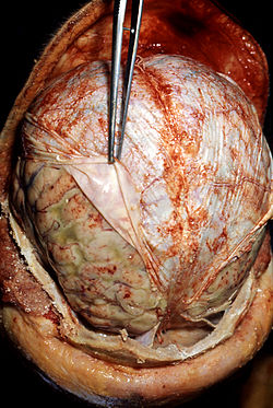
Head opened at autopsy revealing purulent inflammation of leptomeninges beneath reflected dura.
| Beschreibung |
English: Autopsy: Brain surrounded by pus (the yellow-greyish coat around the brain, under the dura lifted by the forceps), the result of bacterial meningitis. A brain autopsy demonstrating signs of pneumococcal meningitis in an alcoholic patient. The forceps (center) are retracting the dura mater (white). Underneath the dura mater are the leptomeninges, which appear to be edematous and have multiple small hemorrhagic foci (red).
Deutsch: Eitrige Haubenmeningitis durch Streptococcus pneumoniae.Autopsie-Befund: unter der zurückgezogenen Dura mater ist die putride Entzündungsreaktion mit kleinen Einblutungen zu erkennen.
Русский: Вскрытие: мозг в окружении гноя (желто-сероватый слой вокруг мозга, оболочка поднимается щипцами), в результате бактериального менингита. Вскрытие мозга демонстрируют признаки менингита. Щипцы (в центре) оттягивают твердую мозговую оболочку (белый). Под твердой мозговой оболочки видны мягкая и паутинная оболочки мозга, которые отёчны и имеют несколько небольших очагов геморрагического воспаления(красный).
Polski: Pneumokokowe zapalenie opon mózgowych. Zdjęcie wykonane podczas sekcji zwłok ukazujące objawy zapalenia opon mózgowo-rdzeniowych wywołanego przez Streptococcus pneumoniae. Pinceta odciąga oponę twardą (biała) pod którą leżą opona pajęcza i opona miękka. Widoczny obrzęk i liczne wybroczyny (czerwone) oraz ropa (zielonkawe). Pneumokokowe zapalenie opon mózgowo-rdzeniowych, treść ropna w przestrzeni podpajeczynówkowej.
Italiano: Autopsia del cervello che mostra un'infezione da meningite pneumococcica in un paziente alcolizzato. Immagine autoptica di encefalo con evidenti depositi di pus dovuto alla presenza di Pneumococco della meningite. La pinza scosta la Dura madre per consentire di visualizzare l'infezione sottostante.
Español: Las zonas de las orillas es el lugar donde ocurre más frecuentemente el sangrado.
Ελληνικά: Αυτοψία: Εγκέφαλος ενδείξεις βακτηριακής μηνιγγίτιδας. |
|||
| Datum | ||||
| Quelle |
|
|||
| Urheber |
|
|||
| Genehmigung (Weiternutzung dieser Datei) |
|
Dateiversionen
Klicke auf einen Zeitpunkt, um diese Version zu laden.
| Version vom | Vorschaubild | Maße | Benutzer | Kommentar | |
|---|---|---|---|---|---|
| aktuell | 23:16, 30. Mär. 2009 | | 1.220 × 1.819 (2,02 MB) | wikimediacommons>Tm | same source |
Dateiverwendung
Die folgende Seite verwendet diese Datei: