Datei:Giardia lamblia SEM 8698 lores.jpg

Größe dieser Vorschau: 509 × 599 Pixel. Weitere Auflösungen: 204 × 240 Pixel | 626 × 737 Pixel.
Originaldatei (626 × 737 Pixel, Dateigröße: 27 KB, MIME-Typ: image/jpeg)
| Beschreibung |
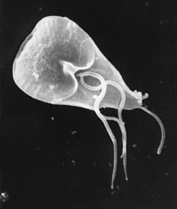
English: This scanning electron micrograph (SEM) revealed some of the external ultrastructural details displayed by a flagellated Giardia lamblia protozoan parasite. G. lamblia is the organism responsible for causing the diarrheal disease "giardiasis". Once an animal or person has been infected with this protozoan, the parasite lives in the intestine, and is passed in the stool. Because the parasite is protected by an outer shell, it can survive outside the body, and in the environment for long periods of time.
Cysts are resistant forms and are responsible for transmission of giardiasis. Both cysts and trophozoites can be found in the feces (diagnostic stages). The cysts are hardy and can survive several months in cold water. Infection occurs by the ingestion of cysts in contaminated water, food, or by the fecal-oral route (hands or fomites). In the small intestine, excystation releases trophozoites (each cyst produces two trophozoites). Trophozoites multiply by longitudinal binary fission, remaining in the lumen of the proximal small bowel where they can be free or attached to the mucosa by a ventral sucking disk. Encystation occurs as the parasites transit toward the colon. The cyst is the stage found most commonly in non-diarrheal feces. Because the cysts are infectious when passed in the stool or shortly afterward, person-to-person transmission is possible. While animals are infected with Giardia, their importance as a reservoir is unclear. |
| Quelle | http://phil.cdc.gov/PHIL_Images/8698/8698_lores.jpg |
| Urheber | CDC / Mahmud Tari |
| Genehmigung (Weiternutzung dieser Datei) |
Copyright Restrictions: None - This image is in the public domain and thus free of any copyright restrictions. As a matter of courtesy we request that the content provider be credited and notified in any public or private usage of this image. |
Dieses Bild ist ein Werk der Centers for Disease Control and Prevention, einer dem Gesundheitsministerium der Vereinigten Staaten unterstellten Behörde, oder es wurde von einem Mitarbeiter dieser Behörde in Ausübung seiner dienstlichen Pflichten erstellt. Als ein Werk der US-amerikanischen Bundesregierung ist dieses Werk in den Vereinigten Staaten gemeinfrei.
|
|
Dieses Medium stammt aus der Public Health Image Library (PHIL), mit der Identifikationsnummer #8698 der Centers for Disease Control and Prevention. Hinweis: Nicht alle PHIL-Bilder sind gemeinfrei; überprüfe unbedingt den Urheberrechtsstatus und die Nennung der Autoren und Inhaltsanbieter.
|
Dateiversionen
Klicke auf einen Zeitpunkt, um diese Version zu laden.
| Version vom | Vorschaubild | Maße | Benutzer | Kommentar | |
|---|---|---|---|---|---|
| aktuell | 13:31, 6. Jul. 2022 | | 626 × 737 (27 KB) | wikimediacommons>Chiswick Chap | crop deadspace |
Dateiverwendung
Die folgenden 2 Seiten verwenden diese Datei: